A new in vivo model using a dorsal skinfold chamber to investigate microcirculation and angiogenesis in diabetic wounds
Stefan Langer 1Christian Beescho 1
Andrej Ring 2
Olivia Dorfmann 1
Hans Ulrich Steinau 3
Nick Spindler 1
1 Department of Plastic, Esthetic and Special Hand Surgery, University Hospital Leipzig, Germany
2 Department of Plastic Surgery and Severe Burns, University Hospital Bergmannsheil, Ruhr University Bochum, Germany
3 Department of Trauma, University Hospital Essen, Germany
Abstract
Introduction: Diabetes mellitus describes a dysregulation of glucose metabolism due to improper insulin secretion, reduced insulin efficacy or both. It is a well-known fact that diabetic patients are likely to suffer from impaired wound healing, as diabetes strongly affects tissue angiogenesis. Until now, no satisfying in vivo murine model has been established to analyze the dynamics of angiogenesis during diabetic wound healing. To help understand the pathophysiology of diabetes and its effect on angiogenesis, a novel in vivo murine model was established using the skinfold chamber in mice.
Materials and Methods: Mutant diabetic mice (db; BKS.Cg-m+/+Leprdb/J), wildtype mice (dock7Leprdb+/+m) and laboratory BALB/c mice were examined. They were kept in single cages with access to laboratory chow with an 12/12 hour day/night circle. Lesions of the panniculus muscle (Ø 2 mm) were created in the center of the transparent window chamber and the subsequent muscular wound healing was then observed for a period of 22 days. Important analytic parameters included vessel diameter, red blood cell velocity, vascular permeability, and leakage of muscle capillaries and post capillary venules. The key parameters were functional capillary density (FCD) and angiogenesis positive area (APA).
Results: We established a model which allows high resolution in vivo imaging of functional angiogenesis in diabetic wounds. As expected, db mice showed impaired wound closure (day 22) compared to wounds of BALB/c or WT mice (day 15). FCD was lower in diabetic mice compared to WT and BALB/c during the entire observation period. The dynamics of angiogenesis also decreased in db mice, as reflected by the lowest APA levels. Significant variations in the skin buildup were observed, with the greatest skin depth in db mice. Furthermore, in db mice, the dermis:subcutaneous ratio was highly shifted towards the subcutaneous layers as opposed to WT or BALB/c mice.
Conclusion: Using this new in vivo model of the skinfold chamber, it was possible to analyze and quantify microangiopathical changes which are essential for a better understanding of the pathophysiology of disturbed wound healing. Research in microcirculation is important to display perfusion in wounds versus healthy tissue. Using our model, we were able to compare wound healing in diabetic and healthy mice. We were also able to objectively analyze perfusion in wound edges and compare microcirculatory parameters. This model may be well suited to augment different therapeutic options.
Keywords
skinfold chamber, microcirculation, angiogenesis, diabetic wounds
Introduction
Diabetes mellitus is defined as a dysregulation of glucose metabolism due to improper insulin secretion, reduced insulin efficacy or both [1]. Diabetes is a common lifelong health condition, which plays an important role in angiogenesis and tissue healing. Tissue healing is a physiological process where reperfusion and angiogenesis of capillaries and soft tissue support wound closure [2]. It is a well-known fact that diabetic patients are likely to suffer from impaired wound healing. Dysregulation in angiogenesis is closely correlated with impaired wound healing.
The capacity to react to hypoxic conditions with adaptive reconstruction of vessels is limited in diabetic patients. When examining the skin of patients with acute hyperinsulinemia (with or without hyperglycemia), no correlation to vessel permeability, hemodynamics, or parameters of endothelial dysfunction could be identified. It remains unclear whether reduced insulin sensitivity is responsible for microcirculatory disturbances [3]. Increased blood viscosity is another pathological factor in diabetics. Moreover, the aggregation of erythrocytes is increased, as is their inability to deform themselves when passing through the capillary system [4], [5], [6], [7], [8]. Unfortunately, there are no in vivo murine models available which would permit close analysis of wound microcirculation in vivo. The aim of the study was to establish a novel mouse model which allows for visualization of microcirculatory disturbances which play a key role in the pathophysiology of diabetic wound healing.
Materials and methods
Diabetic mice from the strain BKS.Cg-m+/+Leprdb/J with a homozygous mutation in the leptin receptor were compared to a control group in terms of macrocirculatory and microcirculatory characteristics. Leptin is an important regulator of the appetite center of the brain, and therefore, diabetic leptin-mutated BKS.Cg-m+/+Leprdb/J mice suffer from polypghagia, polydipsia, and polyuria. After four to eight weeks, the BKS.Cg-m+/+Leprdb/J mice showed increased blood sugar levels.
The control group included wildtype mice of the strain dock7 Leprdb+/+m with the misty mutation. They exhibited normal weight, blood sugar and plasma insulin, had an increased metabolic efficiency, and were non-diabetic. We also used another control group consisting of 8-week-old BALB/c female laboratory mice All mice were weighed at baseline and kept in single housing throughout the experiments.
The model of a transparent skinfold chamber is an established in vivo system that enables studies on angiogenesis. By refining the skinfold chamber through microsurgical implantation techniques, this model could be applied in mice for research purposes. Along with the observation of healthy and pathological tissue it also was possible to examine transplants, tissue replacement substances such as surgical mesh grafts, tissue engineering, and topically/systemically applied medication [9], [10], [11]. The vascularized striated muscle is well represented in the skinfold chamber [12], [13], [14], [15] and can be directly visualized when combined with epifluorescence microscopy.
We implanted the skinfold chamber and created a lesion in the dorsal skin muscle (m. panniculus carnosus) using skin punches (disposable biopsy punch, 2 mm; Stiefel, Germany). A circumscribed circular edge was established and the wound was covered with the coverslip of the skinfold chamber. No air was enclosed and a snap ring fixed the coverslip in place. All mice received a skinfold chamber and the same standardized lesion.
Results
After the chamber implantation, we examined primary wound healing of the intramuscular lesion for a period of 22 days. The mice were immobilized using a PAC (polyacrylate) tube with a frontal valve opening bearing a slit for the skinfold chamber, which was adapted to the exact girth of each mouse. The tube could be fixed on a tablet for microscopic observation.
A digital camera was used to take macroscopic pictures, which served as the basis for the subsequent calculations of the wound area. The microcirculation was documented from 24 hours to 11 days after chamber implantation (see Figure 1 [Fig. 1], Figure 2 [Fig. 2], Figure 3 [Fig. 3]). We acquired additional video data by using 400X magnification to film the edges of the wound and used the computer-assisted image analysis program CapImage (Version 7.4, Dr. Zeintl Software; Heidelberg, Germany). Using this program, we also defined parameters such as vessel diameter (µm), midstream red blood cell velocity (RBCV; mm/s), leakage of muscle capillaries and post capillary venules given by the ratio of fluorescence inside vessel vs outside vessel (Ie/Ii). We also determined the functional capillary density (FCD), meaning red blood cell filled capillaries (mm/mm2).
Figure 1: Hematoxylin and eosin staining of db/db mice (A) and wildtype mice (B). The differences in skin thickness are apparent.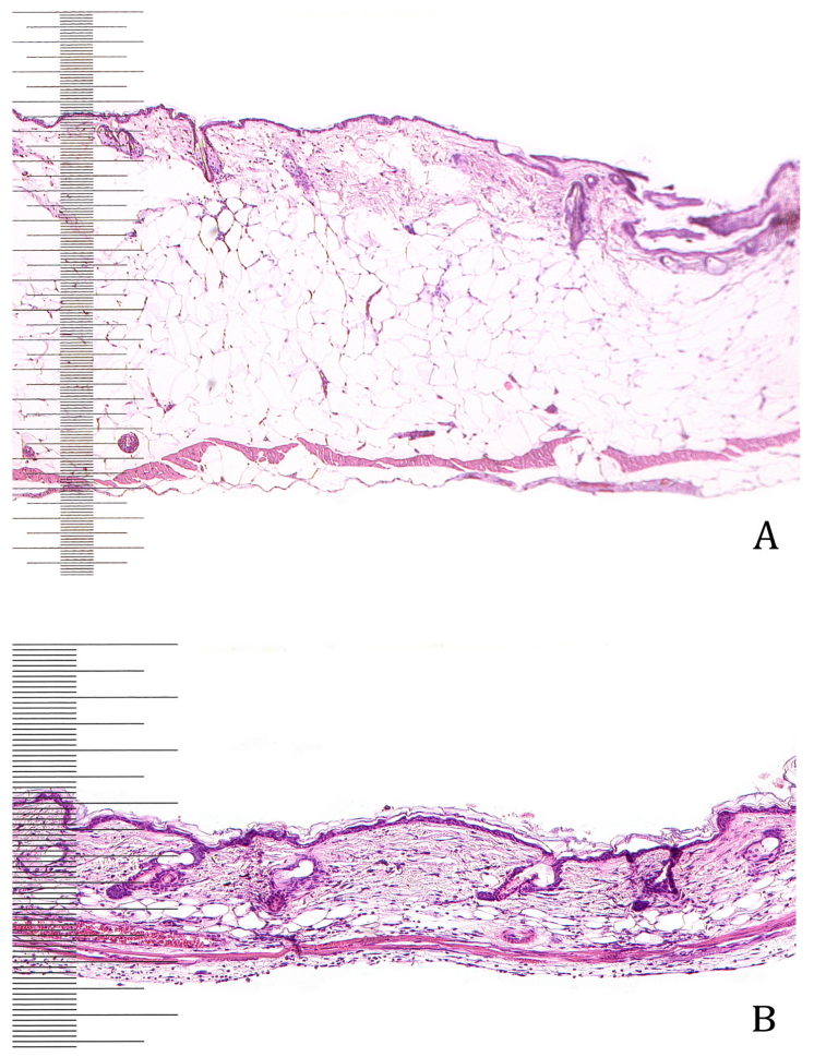
Figure 2: Circular wound of the skin muscle layer on the day of wounding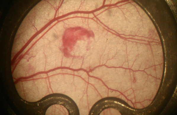
Figure 3: Vessel sprouts visualized using FITC-Dextran as plasma enhancement. Dynamics of vessel development can be analyzed repeatedly over 3 weeks using this model. (bar=10 µm)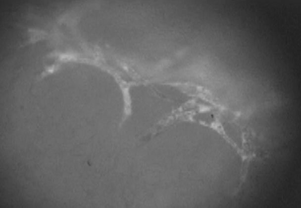
In postcapillary venules, we determined the amount of rolling leucocytes on the endothelium. Angiogenesis positive areas (APA) were counted manually (number/area).
The depth of the dorsal skin varied greatly among our mouse groups. In BALB/c mice, the dorsal skin depth was 370 µm, whereas in dock7Leprdb+/+m mice it was 500 µm. The skin was thickest in db/db mice with values ranging from 1000 to 1400 µm. When comparing the single skin layers among mouse strains, we discovered that the epithelia did not differ significantly. The dermis of db/db mice had a thickness of 250 µm and the subcutaneous layer a thickness of 500 to 1000 µm, providing a dermis: subcutaneous ratio ranging from 1:2 to 1:4. The dermis of BALB/c mice showed a regular and parallel pattern and the thickness was 200 µm. The subcutaneous layer was only 50 µm thick, which leads to a dermis: subcutaneous ratio of 4:1. In dock7Leprdb+/+m mice, the dermis had a thickness of 200 µm, comparable to BALB/c mice; however, the subcutaneous layer was much thicker than in BALB/c mice, with values ranging from 150–200 µm. This resulted in a dermis: subcutaneous ratio of 1:1. The dorsal skin muscle was very compact in wildtype mice, with a thickness ranging from 100 to 150 µm. In BALB/c mice, this muscle was only 30 µm thick. In both control groups, the muscle cells follow a regular, parallel and organized pattern. However, the skin muscle in db/db mice (which is 60 µm thick) shows less compact and poorly organized layers.
Discussion
The skinfold chamber offers the advantage of possible continuous examination and analysis. By fixing the PAC tube on a tablet, it was possible to horizontally position the skinfold chamber for examination using photo- and intravital microscopy. These techniques enabled analysis and quantification of microangiopathical changes, which is essential for understanding the pathophysiology of disturbed wound healing. Research in microcirculation is important to display perfusion in wounds and healthy tissue. Skin perfusion largely depends on the body part. Thus, due to its anatomical localization, striated muscle shows far less fluctuation. Using our in vivo wound healing model, we were able to compare wound healing in diabetic and healthy mice. Our model also enabled us to objectively analyze perfusion within wound edges and compare microcirculatory parameters. In addition, this model offers the opportunity for systemic or topical therapeutic intervention. The influence of extracorporeal shock wave therapy (ECSW) on wound healing has already been successfully examined by our research group. The analytic spectrum will be extended.
Mice make excellent models to study in vivo wound healing, angiogenesis and neovascularization due to their high genomic resemblance to the human genome. BKS.Cg-m+/+Leprdb/J mice are especially suitable for analyzing diabetic wound healing, as they show a homozygous mutation in the leptin receptor. These mice suffer from polypghagia, polydipsia, and polyuria and exhibit the same characteristics as human diabetics. Research conditions can be modified, as different strains of mice may be used to allow the study of genetic variation. In addition to congenitally inbred stems such as BALB/c or C57B1/6 [16], [17], the transparent skinfold chamber can also be used on various genetically modified mice, such as naked mice [18], SCID mice [19], specific knock-out mice [20], [21] or transgenic mice [22]. The skinfold chamber is an ideal model for mice as opposed to other rodents [23], [24].
As mentioned above, we compared coagulation time among different mouse strains during and after surgery while using microsurgical swabs to staunch the flow of blood. Coagulation in BKS.Cg-m+/+Leprdb/J mice was much slower and required more time to be stopped. Only after multiple saline rinses and applying pressure to the tissue was bleeding controlled. Henry et al. confirm that BKS.Cg-m+/+Leprdb/J mice show a prolonged coagulation, which reflects a restricted ADP-dependent thromobocytic aggregation. This is an interesting observation, as type 2 diabetics generally show hypercoagulation associated with myocardial infarcts and stroke due to blood clots in vessels [25].
Prior to implanting the skinfold chamber, the nine week old BKS.Cg-m+/+Leprdb/J mice weighed 36.99 ± 0.91 g, matching the average body weight of 38 g ± SEMxxscanning electron microscope [26]. After the chamber implantation and creation of the wound, the body weight of the mice decreased by 2%. Other studies even describe weight loss of up to 15%, which can be attributed to chamber implantation [27], [28].
Nine-week-old dock7 Leprdb+/+m mice weighed only 24.57 ± 0.59 g. It is also known that high leptin sensitivity keeps animals thin and leptin resistance/lack of leptin leads to obesity. We suspect that the mutation in the leptin receptor in BKS.Cg-m+/+Leprdb/J mice caused the difference in weight of diabetic versus WT mice. Among all groups, BALB/c mice showed the lowest body weight with an average of 19–21 g [29].
Conclusions
The possibilities in research are manifold with the chamber model. Along with the observation of healthy and pathological wound tissue [30], the skinfold chamber offers the tremendous advantage of therapeutic intervention, such as topical or systemic application of medication or application of growth factors (e.g., non viral genetic transfer). Other substances such as transplants, implants and tissue replacement materials may be equally applied [31], [32], [33], [34], [35]. In the past, the skinfold chamber model has been used as a bioreactor for in vivo visualization of transplants or in ischemic reperfusion. Vascularized striated muscle is well represented in the skinfold chamber [36] and can be directly visualized when combined with intra-vital epifluorescence microscopy.
The combination of the skinfold chamber with a transparent window allows continuous observation of the surgical muscle lesion and the daily assessment of wound size. The possibility of intra-vital microscopy is a novelty in a murine model. The chamber model is especially valuable for precise detection of microcirculatory disturbances. To date, no other model comparable to the in vivo skinfold chamber exists and the model presented here is very suitable for long term quantitative analysis of wound healing in diabetic wounds.
Notes
Competing interests
The authors herewith certify that there is no financial or proprietary interest in the subjected matter or materials discussed in this manuscript.
Ethical Standards
Animal studies have been approved by the ethics committee of University of Bochum (AZ 8.87-503709135) and have therefore been performed in accordance with the ethical standards set forth in the 1964 Declaration of Helsinki and its later amendments. The manuscript does not contain clinical studies or patient data.
References
[1] Duraisamy Y, Slevin M, Smith N, Bailey J, Zweit J, Smith C, Ahmed N, Gaffney J. Effect of glycation on basic fibroblast growth factor induced angiogenesis and activation of associated signal transduction pathways in vascular endothelial cells: possible relevance to wound healing in diabetes. Angiogenesis. 2001;4(4):277-88. DOI: 10.1023/A:1016068917266[2] Loots MA, Lamme EN, Mekkes JR, Bos JD, Middelkoop E. Cultured fibroblasts from chronic diabetic wounds on the lower extremity (non-insulin-dependent diabetes mellitus) show disturbed proliferation. Arch Dermatol Res. 1999 Feb-Mar;291(2-3):93-9. DOI: 10.1007/s004030050389
[3] Roszer T. Inflammation as death or life signal in diabetic fracture healing. Inflamm Res. 2011 Jan;60(1):3-10. DOI: 10.1007/s00011-010-0246-9
[4] Mulder DJ, de Boer JF, Graaff R, de Vries R, Annema W, Lefrandt JD, Smit AJ, Tietge UJ, Dullaart RP. Skin autofluorescence is inversely related to HDL anti-oxidative capacity in type 2 diabetes mellitus. Atherosclerosis. 2011 Sep;218(1):102-6. DOI: 10.1016/j.atherosclerosis.2011.05.011
[5] Lerman OZ, Greives MR, Singh SP, Thanik VD, Chang CC, Seiser N, Brown DJ, Knobel D, Schneider RJ, Formenti SC, Saadeh PB, Levine JP. Low-dose radiation augments vasculogenesis signaling through HIF-1-dependent and -independent SDF-1 induction. Blood. 2010 Nov;116(18):3669-76. DOI: 10.1182/blood-2009-03-213629
[6] Patan S. Vasculogenesis and angiogenesis as mechanisms of vascular network formation, growth and remodeling. J Neurooncol. 2000 Oct-Nov;50(1-2):1-15. DOI: 10.1023/A:1006493130855
[7] Buschmann I, Schaper W. Arteriogenesis Versus Angiogenesis: Two Mechanisms of Vessel Growth. News Physiol Sci. 1999 Jun;14:121-125.
[8] Shiekh GA, Ayub T, Khan SN, Dar R, Andrabi KI. Reduced nitrate level in individuals with hypertension and diabetes. J Cardiovasc Dis Res. 2011 Jul;2(3):172-6. DOI: 10.4103/0975-3583.85264
[9] Graier WF, Posch K, Wascher TC, Kostner GM. Role of superoxide anions in changes of endothelial vasoactive response during acute hyperglycemia. Horm Metab Res. 1997 Dec;29(12):622-6. DOI: 10.1055/s-2007-979113
[10] Zoppini G, Targher G, Cacciatori V, Guerriero A, Muggeo M. Chronic cigarette smoking is associated with increased plasma circulating intercellular adhesion molecule 1 levels in young type 1 diabetic patients. Diabetes Care. 1999 Nov;22(11):1871-4. DOI: 10.2337/diacare.22.11.1871
[11] Taguchi K, Kobayashi T, Takenouchi Y, Matsumoto T, Kamata K. Angiotensin II causes endothelial dysfunction via the GRK2/Akt/eNOS pathway in aortas from a murine type 2 diabetic model. Pharmacol Res. 2011 Nov;64(5):535-46. DOI: 10.1016/j.phrs.2011.05.001
[12] Ring A, Goertz O, Muhr G, Steinau HU, Langer S. In vivo microvascular response of murine cutaneous muscle to ibuprofen-releasing polyurethane foam. Int Wound J. 2008 Jun;5(3):464-9. DOI: 10.1111/j.1742-481X.2007.00403.x
[13] Ring A, Mullershausen F, Goertz O, Koesling D, Muhr G, Steinau HU, Steinstraesser L, Langer S. Exogenous nitric oxide donation causes desensitization of arteriolar relaxing activity in vivo: an intravital analysis in mice. J Surg Res. 2010 Nov;164(1):169-74. DOI: 10.1016/j.jss.2009.07.032
[14] Morise T, Takeuchi Y, Kawano M, Koni I, Takeda R. Increased plasma levels of immunoreactive endothelin and von Willebrand factor in NIDDM patients. Diabetes Care. 1995 Jan;18(1):87-9.
[15] Lindenblatt N, Platz U, Althaus M, Hegland N, Schmidt CA, Contaldo C, Vollmar B, Giovanoli P, Calcagni M. Temporary angiogenic transformation of the skin graft vasculature after reperfusion. Plast Reconstr Surg. 2010 Jul;126(1):61-70. DOI: 10.1097/PRS.0b013e3181da87f6
[16] Dedeugd C, Wankhede M, Sorg BS. Multimodal optical imaging of microvessel network convective oxygen transport dynamics. Appl Opt. 2009 Apr;48(10):D187-97. DOI: 10.1364/ao.48.00d187
[17] Isaka N, Padera TP, Hagendoorn J, Fukumura D, Jain RK. Peritumor lymphatics induced by vascular endothelial growth factor-C exhibit abnormal function. Cancer Res. 2004 Jul;64(13):4400-4. DOI: 10.1158/0008-5472.CAN-04-0752
[18] Fiedler U, Reiss Y, Scharpfenecker M, Grunow V, Koidl S, Thurston G, Gale NW, Witzenrath M, Rosseau S, Suttorp N, Sobke A, Herrmann M, Preissner KT, Vajkoczy P, Augustin HG. Angiopoietin-2 sensitizes endothelial cells to TNF-alpha and has a crucial role in the induction of inflammation. Nat Med. 2006 Feb;12(2):235-9. DOI: 10.1038/nm1351
[19] Li H, Hergert SM, Schäfer SC, Brausch I, Yao Y, Huang Q, Mang C, Lehr HA, Förstermann U. Midostaurin upregulates eNOS gene expression and preserves eNOS function in the microcirculation of the mouse. Nitric Oxide. 2005 Jun;12(4):231-6. DOI: 10.1016/j.niox.2005.04.001
[20] Frost GI, Lustgarten J, Dudouet B, Nyberg L, Hartley-Asp B, Borgström P. Novel syngeneic pseudo-orthotopic prostate cancer model: vascular, mitotic and apoptotic responses to castration. Microvasc Res. 2005 Jan;69(1-2):1-9. DOI: 10.1016/j.mvr.2004.10.001
[21] Goncalves AC, Tank J, Diedrich A, Hilzendeger A, Plehm R, Bader M, Luft FC, Jordan J, Gross V. Diabetic hypertensive leptin receptor-deficient db/db mice develop cardioregulatory autonomic dysfunction. Hypertension. 2009 Feb;53(2):387-92. DOI: 10.1161/HYPERTENSIONAHA.108.124776
[22] Kieffer TJ, Heller RS, Habener JF. Leptin receptors expressed on pancreatic beta-cells. Biochem Biophys Res Commun. 1996 Jul;224(2):522-7. DOI: 10.1006/bbrc.1996.1059
[23] Henry ML, Davidson LB, Wilson JE, McKenna BK, Scott SA, McDonagh PF, Ritter LS. Whole blood aggregation and coagulation in db/db and ob/ob mouse models of type 2 diabetes. Blood Coagul Fibrinolysis. 2008 Mar;19(2):124-34. DOI: 10.1097/MBC.0b013e3282f41e56
[24] Acang N, Jalil FD. Hypercoagulation in diabetes mellitus. Southeast Asian J Trop Med Public Health. 1993;24 Suppl 1:263-6.
[25] Crijns FR, Struijker Boudier HA, Wolffenbuttel BH. Arteriolar reactivity in conscious diabetic rats: influence of aminoguanidine treatment. Diabetes. 1998 Jun;47(6):918-23. DOI: 10.2337/diabetes.47.6.918
[26] Leunig M, Yuan F, Menger MD, Boucher Y, Goetz AE, Messmer K, Jain RK. Angiogenesis, microvascular architecture, microhemodynamics, and interstitial fluid pressure during early growth of human adenocarcinoma LS174T in SCID mice. Cancer Res. 1992 Dec;52(23):6553-60.
[27] Pico C, Jilkova ZM, Kus V, Palou A, Kopecky J. Perinatal programming of body weight control by leptin: putative roles of AMP kinase and muscle thermogenesis. Am J Clin Nutr. 2011 Dec;94(6 Suppl):1830S-1837S. DOI: 10.3945/ajcn.110.000752
[28] Yamada R, Horinouch H, Tsukada K. Spatiotemporal measurement of tumor oxygenation reveals repeat hypoxic phenomenon in mice. Conf Proc IEEE Eng Med Biol Soc. 2011;2011:5965-8. DOI: 10.1109/IEMBS.2011.6091474
[29] Upreti M, Jamshidi-Parsian A, Koonce NA, Webber JS, Sharma SK, Asea AA, Mader MJ, Griffin RJ. Tumor-Endothelial Cell Three-dimensional Spheroids: New Aspects to Enhance Radiation and Drug Therapeutics. Transl Oncol. 2011 Dec;4(6):365-76. DOI: 10.1593/tlo.11187
[30] Laschke MW, Vollmar B, Menger MD. The dorsal skinfold chamber: window into the dynamic interaction of biomaterials with their surrounding host tissue. Eur Cell Mater. 2011;22:147-64; discussion 164-7.
[31] Kraft CN, Burian B, Perlick L, Wimmer MA, Wallny T, Schmitt O, Diedrich O. Impact of a nickel-reduced stainless steel implant on striated muscle microcirculation: a comparative in vivo study. J Biomed Mater Res. 2001 Dec;57(3):404-12. DOI: 10.1002/1097-4636(20011205)57:3<404::AID-JBM1183>3.0.CO;2-W
[32] Rezaeian F, Wettstein R, Scheuer C, Bäumker K, Bächle A, Vollmar B, Menger MD, Harder Y. Ghrelin protects musculocutaneous tissue from ischemic necrosis by improving microvascular perfusion. Am J Physiol Heart Circ Physiol. 2012 Feb;302(3):H603-10. DOI: 10.1152/ajpheart.00390.2010
[33] Nishimura R, Goto M, Sekiguchi S, Fujimori K, Ushiyama A, Satomi S. Assessment for revascularization of transplanted pancreatic islets at subcutaneous site in mice with a highly sensitive imaging system. Transplant Proc. 2011 Nov;43(9):3239-40. DOI: 10.1016/j.transproceed.2011.09.095
[34] Menger MD, Barker JH, Messmer K. Capillary blood perfusion during postischemic reperfusion in striated muscle. Plast Reconstr Surg. 1992 Jun;89(6):1104-14. DOI: 10.1097/00006534-199206000-00017
[35] Nolte D, Bayer M, Lehr HA, Becker M, Krombach F, Kreimeier U, Messmer K. Attenuation of postischemic microvascular disturbances in striated muscle by hyperosmolar saline dextran. Am J Physiol. 1992 Nov;263(5 Pt 2):H1411-6.
[36] Sirsjö A, Lehr HA, Nolte D, Haapaniemi T, Lewis DH, Nylander G, Messmer K. Hyperbaric oxygen treatment enhances the recovery of blood flow and functional capillary density in postischemic striated muscle. Circ Shock. 1993 May;40(1):9-13.

